| Cat # | Size | Price | Quantity | |
|---|---|---|---|---|
| 109901 | 100 ug | $80 | ||
| 109902 | 500 ug | $220 |
| Clone | OKT11 |
|---|---|
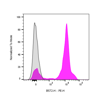
| Application | Flow Cytometry |
| Reactivity | Human |
| Format | Purified |
| Target Name | CD2, LFA-2, SRBC-R |
| Isotype | Mouse IgG1 |
| Antibody Type | Monoclonal |
| Regulatory Status | RUO |
| Formulation | Phosphate-buffered solution, pH 7.2, containing 0.09% sodium azide |
| Protein Concentration | 0.5 mg/mL |
| Storage&Handling | The antibody solution should be stored between 2°C and 8°C |
| Recommended Usage | For flow cytometric staining, it is recommended to use less than 0.25 µg of this reagent per 0.5-1.0 million cells in a 100 µL volume. Optimal reagent performance should be determined by titration for each specific application |
| See All Formats | Clone OKT11 |
CD2 is a transmembrane glycoprotein expressed primarily on T lymphocytes and natural killer (NK) cells, where it plays an essential role in cell adhesion, activation, and immune recognition. It is one of the earliest identified surface markers of T cells and serves as a critical component of the immunological synapse between T cells and antigen-presenting cells (APCs). Through its interactions, CD2 enhances the efficiency of antigen-dependent T cell activation and mediates stable cell–cell contact necessary for effective immune signaling.
Structurally, CD2 is a type I transmembrane protein of approximately 50 kDa, consisting of a short cytoplasmic tail, a single hydrophobic transmembrane region, and an extracellular segment composed of two immunoglobulin-like domains. These domains are responsible for binding to specific ligands on target cells. The cytoplasmic region contains several sites that interact with intracellular signaling and cytoskeletal molecules, allowing CD2 to couple cell adhesion with activation pathways. This structural arrangement enables CD2 to act as both an adhesion molecule and a co-stimulatory receptor.
The primary ligands for CD2 are CD58 (LFA-3) in humans and CD48 in rodents. The CD2-CD58 interaction is of high affinity and stabilizes contact between T cells and antigen-presenting cells, facilitating T cell receptor (TCR) signaling and cytokine production. Beyond adhesion, CD2 engagement modulates calcium flux, cytoskeletal rearrangement, and the expression of activation markers, contributing to T cell proliferation and cytotoxicity. On NK cells, CD2 enhances target recognition and the release of cytolytic granules.
Dysregulation of CD2 expression or signaling has been associated with immune disorders and malignancy. In autoimmune diseases such as multiple sclerosis and rheumatoid arthritis, excessive T cell activation involves augmented CD2-CD58 interactions. Altered CD2 expression patterns are also noted in certain leukemias and lymphomas, including T-cell acute lymphoblastic leukemia, where it serves as a diagnostic marker. In viral infections, manipulation of CD2-mediated adhesion can modulate immune evasion mechanisms.
Therapeutically, CD2 represents a target for immune modulation. Monoclonal antibodies directed against CD2, such as alefacept, have been developed to suppress pathogenic T cell activity in autoimmune diseases like psoriasis. Modulation of CD2-CD58 interactions also has potential applications in transplantation tolerance and cancer immunotherapy. By regulating T cell activation and adhesion, therapeutic targeting of CD2 offers a means to rebalance immune responses across a spectrum of inflammatory and malignant disorders.
Mouse IgG1 Isotype Control Antibody
Mouse IgG1 Isotype Control Antibody
Have a product or application question? Consult our FAQs or contact us.