| Cat # | Size | Price | Quantity | |
|---|---|---|---|---|
| 202701 | 100 ug | $90 | ||
| 202702 | 25 tests | $85 |
| Clone | DX5_R |
|---|---|
| Application | Flow Cytometry |
| Reactivity | Mouse |
| Format | Purified |
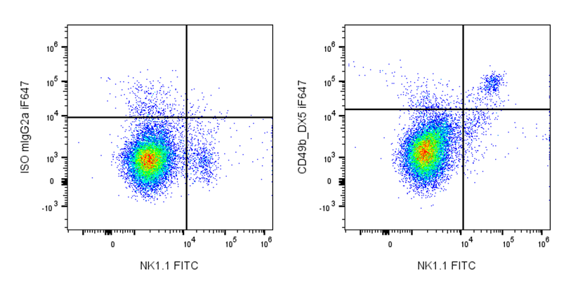
| Target Name | CD49b, Integrin alpha 2 chain, ITGA2 |
| Isotype | Mouse IgG2a IgM like |
| Antibody Type | Monoclonal |
| Regulatory Status | RUO |
| Formulation | Phosphate-buffered solution, pH 7.2, containing 0.09% sodium azide |
| Protein Concentration | 0.5 mg/mL |
| Storage&Handling | The antibody solution should be stored between 2°C and 8°C |
| Recommended Usage | For flow cytometric staining, it is recommended to use less than 0.2 µg of this reagent per 0.5-1.0 million cells in a 100 µL volume. Optimal reagent performance should be determined by titration for each specific application. |
| See All Formats | Clone DX5_R |
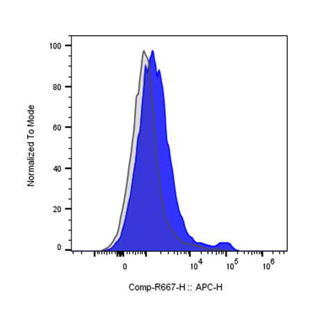
Mouse CD49b, also known as integrin α2 (Itga2), is a cell surface adhesion receptor best known in immunology as a defining marker of natural killer (NK) cells in mice. CD49b pairs with the integrin β1 subunit (CD29) to form the α2β1 integrin, also referred to as very late antigen-2 (VLA-2). Through this heterodimeric complex, CD49b mediates cell adhesion, migration, and signaling, supporting immune cell interactions with the extracellular matrix and other cells.
Structurally, CD49b is a type I transmembrane glycoprotein with a large extracellular domain, a single transmembrane helix, and a short cytoplasmic tail. The extracellular region contains an inserted (I) domain, also called an A domain, which is responsible for ligand binding and requires divalent cations such as Mg2+ or Mn2+ for activity. Like other integrins, CD49b undergoes conformational changes that regulate ligand affinity and enable bidirectional “inside-out” and “outside-in” signaling. The cytoplasmic tail lacks intrinsic enzymatic activity but associates with adaptor proteins that link the receptor to the actin cytoskeleton.
The primary ligands for mouse CD49b are extracellular matrix proteins, most notably collagen types I, II, and IV, as well as laminin. Binding to these ligands enables NK cells and other CD49b-expressing cells to adhere to tissue matrices and migrate within peripheral tissues. Through α2β1 integrin signaling, CD49b contributes to cellular activation, survival, and cytotoxic function, particularly in tissue-resident or tissue-infiltrating immune populations.
CD49b has important implications in disease and immune regulation. In mouse models, CD49b expression is widely used to identify and study NK cells and NKT cell subsets, including in cancer, infection, and autoimmune disease research. Altered α2β1 integrin signaling has been linked to dysregulated immune cell trafficking, chronic inflammation, and fibrotic processes. In cancer models, CD49b-positive NK cells play a key role in antitumor immunity through their ability to localize to tumor tissues and mediate cytotoxic responses.
Therapeutically, mouse CD49b is primarily leveraged as a biomarker and experimental targeting handle rather than a direct clinical target. Antibodies against CD49b (such as DX5) are widely used to identify, isolate, or deplete NK cells in preclinical studies. Insights gained from CD49b-defined NK cell biology inform the development of NK cell–based immunotherapies and strategies aimed at enhancing immune cell trafficking and function, underscoring the translational relevance of this integrin in immunology research.
The HMα2 antibody has been shown to be useful for partially blocking CD49b mediated interactions with collagen. Additionally, this antibody blocks staining of splenic NK cells by the monoclonal antibody DX5.
Mouse IgG2a Isotype Control Antibody
Anti-Mouse CD49b (pan-NK, integrin α2) Antibody TDS
Mouse IgG2a Isotype Control Antibody
Have a product or application question? Consult our FAQs or contact us.