| Cat # | Size | Price | Quantity | |
|---|---|---|---|---|
| 108303 | 25 tests | $30 | ||
| 108304 | 100 tests | $60 |
| Clone | RPA-T8 |
|---|---|
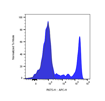
| Application | Flow Cytometry |
| Reactivity | Human |
| Format | APC |
| Target Name | CD8, T8, Leu2 |
| Isotype | Mouse IgG1 |
| Antibody Type | Monoclonal |
| Regulatory Status | RUO |
| Formulation | Phosphate-buffered solution, pH 7.2, containing 0.09% sodium azide and 0.2% (w/v) BSA |
| Protein Concentration | Supplied at a lot-specific concentration. |
| Storage&Handling | The antibody solution should be stored undiluted between 2°C and 8°C, and protected from prolonged exposure to light. Do not freeze. |
| Recommended Usage | For flow cytometric staining, it is recommended to use 5 µL of this reagent per 0.5-1.0 million cells in a 100 µL volume. Optimal reagent performance should be determined by titration for each specific application. APC has an excitation max at 650 nm and an emission max at 660 nm. |
| Excitation Laser | Red Laser (633 nm) |
| See All Formats | Clone RPA-T8 |
CD8 is a cell surface glycoprotein that plays a critical role in cellular immunity. It is most commonly expressed on cytotoxic T lymphocytes (CD8⁺ T cells), although lower levels can also be found on subsets of natural killer (NK) cells and dendritic cells.
Structurally, CD8 exists either as a homodimer of two CD8α chains (CD8αα) or, more commonly on T cells, as a heterodimer composed of CD8α and CD8β chains (CD8αβ). Each chain contains an extracellular immunoglobulin-like domain, a transmembrane region, and a short cytoplasmic tail that associates with intracellular signaling molecules. Functionally, CD8 acts as a co-receptor for the T cell receptor (TCR) during antigen recognition. Its primary ligand is major histocompatibility complex class I (MHC I), which is expressed on nearly all nucleated cells. During immune surveillance, the TCR recognizes peptide antigens presented by MHC I, while CD8 binds to a conserved region of the MHC I molecule. This interaction stabilizes the TCR-peptide-MHC complex and brings the Src-family kinase Lck into proximity with the TCR signaling machinery, thereby enhancing signal transduction and lowering the threshold for T cell activation. Once activated, CD8⁺ T cells differentiate into cytotoxic effector cells capable of directly killing infected or malignant cells. They mediate target cell death primarily through the release of perforin and granzymes, which induce apoptosis, as well as through engagement of death receptor pathways such as Fas-Fas ligand interactions. CD8⁺ T cells also secrete cytokines, including interferon-γ (IFN-γ) and tumor necrosis factor-α (TNF-α), which further shape immune responses and inhibit pathogen replication.
CD8⁺ T cells are essential for protection against viral infections and for immune surveillance against cancer. However, their dysregulation contributes to disease. Insufficient CD8⁺ T cell responses can result in chronic viral infections or tumor immune evasion, while excessive or misdirected activity can cause tissue damage and contribute to autoimmune diseases, such as type 1 diabetes and multiple sclerosis. In chronic infections and cancer, persistent antigen exposure can drive CD8⁺ T cell exhaustion, characterized by reduced effector function and sustained expression of inhibitory receptors.
In therapeutics, CD8⁺ T cells are central to modern immunotherapy. Cancer treatments such as immune checkpoint inhibitors aim to reinvigorate exhausted CD8⁺ T cells, while adoptive cell therapies, including CAR-T and TCR-engineered T cells, harness or enhance CD8⁺ cytotoxic activity. CD8⁺ T cell responses are also a key goal of antiviral vaccines, underscoring their importance in both disease control and therapeutic intervention.
APC Mouse IgG1 Isotype Control Antibody
APC Anti-Human CD8a Antibody TDS
APC Mouse IgG1 Isotype Control Antibody
Have a product or application question? Consult our FAQs or contact us.