| Cat # | Size | Price | Quantity | |
|---|---|---|---|---|
| 100105 | 25 tests | $95 | ||
| 100106 | 100 tests | $240 |
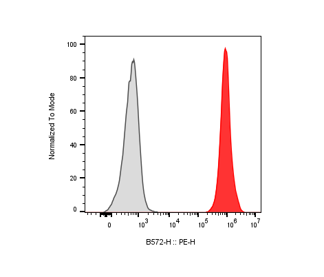
| Clone | 528 |
|---|---|
| Application | Flow Cytometry |
| Reactivity | Human |
| Format | PE |
| Target Name | EGFR, Proto-oncogene c-ErbB-1, Receptor tyrosine-protein kinase erbB-1, HER1 |
| Isotype | Mouse IgG2a |
| Antibody Type | Monoclonal |
| Regulatory Status | RUO |
| Formulation | Phosphate-buffered solution, pH 7.2, containing 0.09% sodium azide and 0.2% (w/v) BSA |
| Protein Concentration | Supplied at a lot-specific concentration. |
| Storage&Handling | The antibody solution should be stored undiluted between 2°C and 8°C, and protected from prolonged exposure to light. Do not freeze. |
| Recommended Usage | For flow cytometric staining, it is recommended to use 5 µL of this reagent per 0.5-1.0 million cells in a 100 µL volume. Optimal reagent performance should be determined by titration for each specific application. PE has an excitation max at 565 nm and an emission max at 575 nm. |
| Excitation Laser | Blue Laser (488 nm) Green/Yellow laser (532/561nm) |
| URL | https://dev.innocyto.com/web/pe-anti-human-egfr-2361 |
| See All Formats | Clone 528 |
The epidermal growth factor receptor (EGFR), also known as ErbB1 or HER1, is a transmembrane receptor tyrosine kinase that plays a central role in regulating cell growth, survival, differentiation, and migration. EGFR is a member of the ErbB family of receptors and is expressed in many epithelial, mesenchymal, and neural tissues.
Under normal physiological conditions, EGFR signaling is tightly controlled and essential for tissue development, maintenance, and repair. EGFR is activated through the binding of specific ligands to its extracellular domain. Key ligands include epidermal growth factor (EGF), transforming growth factor-α (TGF-α), amphiregulin, epiregulin, betacellulin, and heparin-binding EGF-like growth factor (HB-EGF). Ligand binding induces receptor dimerization, either as EGFR homodimers or heterodimers with other ErbB family members such as ErbB2 (HER2). Dimerization triggers autophosphorylation of tyrosine residues within the intracellular kinase domain, creating docking sites for adaptor proteins and initiating downstream signaling cascades. Major pathways activated by EGFR include the MAPK/ERK, PI3K/AKT, and JAK/STAT pathways, which collectively regulate proliferation, survival, and gene expression.
Dysregulation of EGFR signaling is strongly associated with human disease, particularly cancer. EGFR can become aberrantly activated through overexpression, gene amplification, activating mutations, or autocrine ligand production. Such alterations drive uncontrolled cell proliferation and resistance to apoptosis. EGFR dysregulation is commonly observed in cancers of the lung, colorectal tract, head and neck, pancreas, and glioblastoma. Specific activating mutations in EGFR, such as exon 19 deletions or the L858R point mutation, are especially prevalent in subsets of non-small cell lung cancer and are associated with sensitivity to targeted therapies. Beyond cancer, EGFR signaling has also been implicated in inflammatory diseases, tissue fibrosis, and abnormal wound healing.
Because of its central role in disease pathogenesis, EGFR is an important therapeutic target. Multiple classes of EGFR-targeted therapies have been developed, including small-molecule tyrosine kinase inhibitors (TKIs) and monoclonal antibodies that block ligand binding or receptor dimerization. EGFR inhibitors have demonstrated significant clinical benefit in selected patient populations, particularly those with EGFR-driven tumors, making EGFR one of the most extensively studied and successfully targeted receptors in modern precision medicine.
PE Mouse IgG2a Isotype Control Antibody
PE Anti-Human EGFR Antibody TDS
PE Mouse IgG2a Isotype Control Antibody
Have a product or application question? Consult our FAQs or contact us.