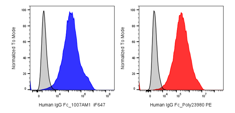
| Cat # | Size | Price | Quantity | |
|---|---|---|---|---|
| 306601 | 25 µg | $80 | ||
| 306602 | 100 µg | $180 |
| Clone | 1007AM1 |
|---|---|
| Application | Flow Cytometry |
| Reactivity | Human IgG Fcγ |
| Format | iF647 |
| Target Name | Human IgG Fcγ |
| Isotype | Mouse IgG1 |
| Antibody Type | Monoclonal |
| Regulatory Status | RUO |
| Formulation | Phosphate-buffered solution, pH 7.2, containing 0.09% sodium azide and 0.2% (w/v) BSA |
| Protein Concentration | 0.2 mg/mL |
| Storage&Handling | The antibody solution should be stored undiluted between 2°C and 8°C, and protected from prolonged exposure to light. Do not freeze. |
| Recommended Usage | For flow cytometric staining, it is recommended to use less than 0.2 ug of this reagent per 0.5-1.0 million cells in a 100 µL volume. Optimal reagent performance should be determined by titration for each specific application |
| Excitation Laser | Red Laser (633 nm) |
| See All Formats | Clone 1007AM1 |
The human IgG Fc region is the constant (fragment crystallizable) portion of immunoglobulin G (IgG) antibodies that mediates communication between antibodies and the immune system. Although the Fc region does not bind antigen directly, it recruits immune effector mechanisms after the antigen-binding Fab regions recognize their targets. Through interactions with Fc gamma (Fcγ) receptors expressed on macrophages, neutrophils, dendritic cells, natural killer (NK) cells, and other leukocytes, as well as complement component C1q, the IgG Fc region triggers antibody-dependent cellular cytotoxicity (ADCC), antibody-dependent cellular phagocytosis (ADCP), complement-dependent cytotoxicity (CDC), and modulation of inflammatory responses. The Fc region also binds the neonatal Fc receptor (FcRn), which protects IgG from lysosomal degradation and extends its serum half-life to approximately three weeks.
Structurally, the IgG Fc region consists of the paired heavy-chain constant domains CH2 and CH3 connected by a flexible hinge region. The Fc forms a homodimer stabilized by disulfide bonds and contains a conserved N-linked glycosylation site at Asn297 within each CH2 domain. Fc glycosylation is essential for maintaining the proper conformation required for Fcγ receptor and C1q binding, and changes in glycan composition can markedly alter antibody effector function. Major ligands include activating Fcγ receptors (FcγRI, FcγRIIA, FcγRIIIA), the inhibitory receptor FcγRIIB, FcRn, and C1q.
Altered Fc-mediated signaling contributes to numerous diseases. Excessive Fcγ receptor activation by immune complexes promotes autoimmune disorders such as systemic lupus erythematosus, rheumatoid arthritis, and immune thrombocytopenia, while impaired Fc function can reduce pathogen clearance and antitumor immunity. Differences in Fc glycosylation have also been associated with chronic inflammatory diseases, infections, and cancer.
The IgG Fc region is a major focus of therapeutic antibody engineering. Fc modifications can enhance or reduce Fcγ receptor binding, prolong serum half-life through improved FcRn interactions, or eliminate unwanted effector functions. Engineered Fc domains are widely incorporated into monoclonal antibodies, bispecific antibodies, Fc-fusion proteins, and antibody-drug conjugates to optimize efficacy, safety, and pharmacokinetics, making the human IgG Fc region a cornerstone of modern immunotherapy.
iF647 Anti-Human IgG Fc antibody TDS
Have a product or application question? Consult our FAQs or contact us.