Flow cytometry has become the workhorse technique for immunophenotyping, functional assessment, and biomarker discovery across immunology and oncology research. Yet even the most carefully designed panel can be undermined by a persistent and often underestimated problem: background signal. Whether it arises from nonspecific antibody binding, autofluorescence, or improper compensation, background noise erodes the very thing flow cytometry is meant to deliver — a clean, quantifiable readout of biological truth. Understanding where background comes from, and how to systematically eliminate it, is essential for generating data that holds up to peer review and downstream decision-making.
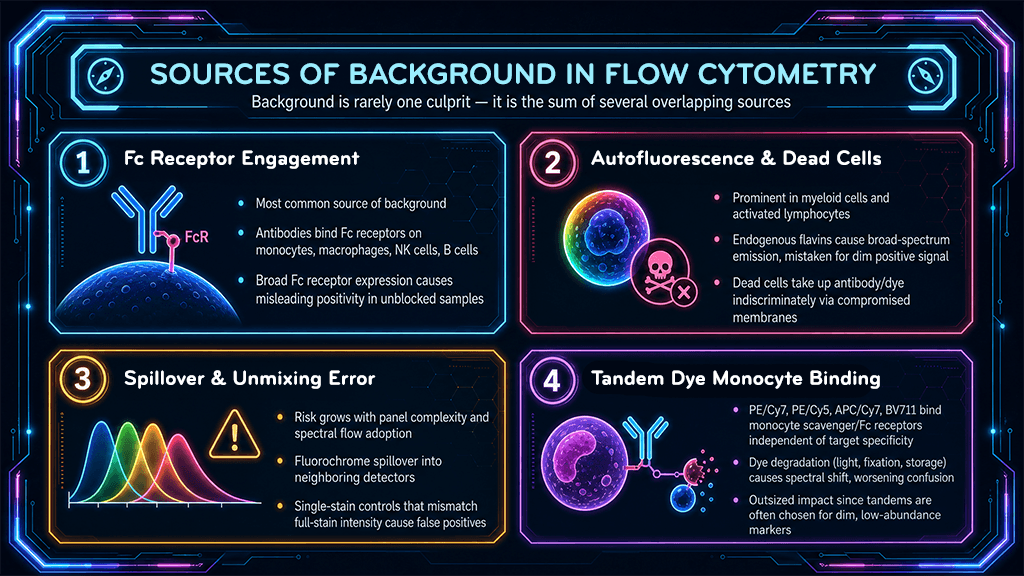
Background in flow cytometry is rarely a single culprit; it is the sum of several overlapping sources. Nonspecific antibody binding is the most common offender, occurring when antibodies attach to Fc receptors on monocytes, macrophages, NK cells, and B cells rather than to their intended target antigen. Because Fc receptors are expressed broadly across immune cell subsets, unblocked samples can show elevated and misleading positivity in populations that never truly engaged the antibody's target.
Autofluorescence is a second major contributor, particularly in myeloid cells, activated lymphocytes, and samples containing dead or dying cells. Granulocytes and monocytes naturally emit fluorescence across a broad spectral range due to endogenous flavins and other metabolic byproducts, which can be easily mistaken for a true positive signal in dim channels. Dead cells compound this problem further, as compromised membranes allow antibodies and fluorochromes to enter indiscriminately, producing a diffuse, unspecific signal that inflates apparent marker expression.
A third source is spectral overlap and poor compensation. As panels grow in complexity (a trend accelerating with the adoption of spectral flow cytometry), the risk of fluorochrome spillover into neighboring detectors increases. Inadequate compensation or unmixing can manifest as false-positive events in channels where no true signal exists, particularly when single-stain controls do not accurately represent the fluorescence intensity of the fully stained sample.
Tandem dyes, such as PE/Cyanine7, PE/Cyanine5, APC/Cyanine7, and BV711, introduce a background problem distinct from simple nonspecific binding: monocytes have a well-documented tendency to bind these polymer-based and cyanine-linked conjugates independent of the antibody's target specificity. This occurs because monocytes express scavenger receptors and Fc receptors that recognize structural features of the tandem fluorochrome itself, and in some cases the dye backbone can be taken up via nonspecific endocytic or membrane-adsorption mechanisms. The result is a false-positive signal that appears specifically in the monocyte gate, regardless of antibody specificity, resulting in a pattern that can easily be mistaken for genuine marker expression if the analyst is not specifically looking for it. This issue is compounded by tandem dye degradation, where the acceptor fluorophore separates from the donor over time (due to light exposure, fixation, or prolonged storage), causing spectral shifts that further confound compensation and make the nonspecific monocyte signal harder to distinguish from true positivity in neighboring channels. Because tandem dyes are frequently chosen for dim or low- abundance markers in crowded panels, this artifact has an outsized impact on data interpretation precisely where sensitivity matters most.
Uncontrolled background does more than add noise; it actively distorts biological interpretation. Rare population analysis, such as identifying antigen-specific T cells, minimal residual disease, or low-frequency stem and progenitor populations, is especially vulnerable, since even a small percentage of nonspecific events can overwhelm a population that represents less than 0.1% of total cells. Gating strategies built on noisy data can shift population boundaries, leading to inconsistent results between operators, instruments, and labs.
This is not merely an academic concern. In translational and clinical research, background-driven misclassification can affect patient stratification, therapeutic response assessment, and reproducibility across multi-site trials. As flow cytometry panels expand toward 20-, 30-, and even 40-plus color designs, the tolerance for background error shrinks correspondingly, making rigorous background control a prerequisite rather than an optional refinement.
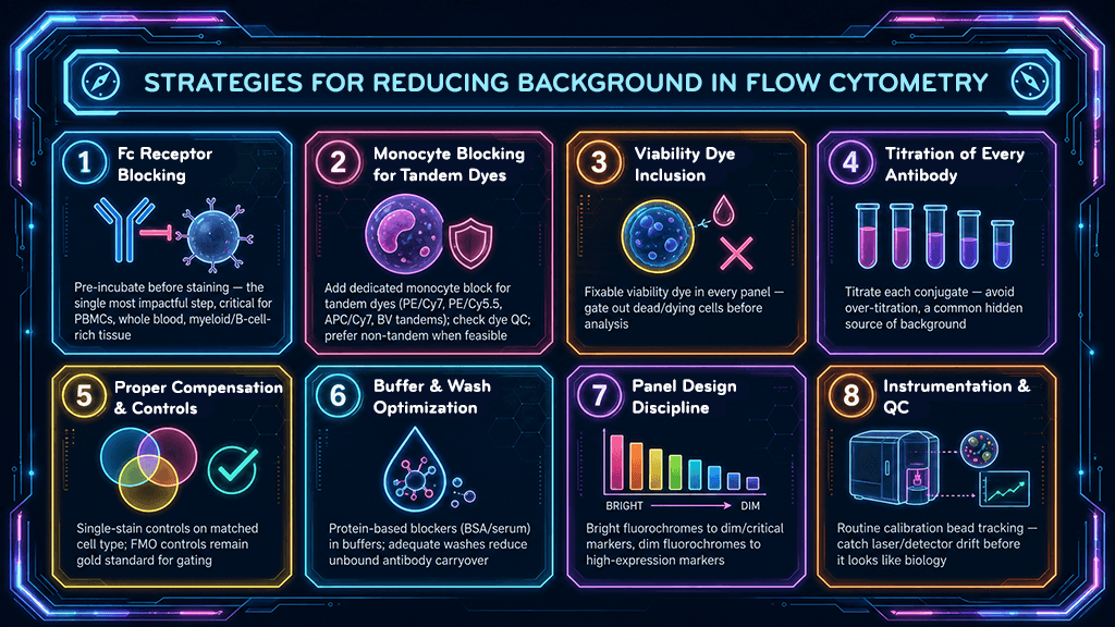
Fc Receptor Blocking. The single most impactful step for reducing nonspecific antibody binding is pre-incubation with an Fc receptor blocking reagent prior to antibody staining. This step is particularly critical when working with human PBMCs, whole blood, or tissues rich in myeloid and B cell populations, where Fc receptor density is high. Skipping this step is one of the most common — and most easily corrected — sources of inflated background in published flow data.
Monocyte Blocking for Tandem Dye Conjugates. Standard Fc receptor blocking reagents do not fully address nonspecific tandem dye uptake by monocytes, since this binding is mediated by scavenger receptor and membrane interactions rather than classical Fc receptor engagement alone. A dedicated monocyte blocking reagent, used in addition to standard Fc block, substantially reduces this artifact and should be considered mandatory in any panel using tandem dyes such as PE/Cyanine7, PE/Cyanine5.5, APC/Cyanine7, or polymer-based Brilliant Violet tandems. Alongside blocking, verifying tandem dye integrity before each experiment, by checking lot-specific QC data or running a brief single-stain check for degradation, helps confirm that any monocyte-associated signal reflects true biology rather than dye breakdown. Where feasible, substituting a non-tandem fluorochrome for markers expressed on monocyte populations of interest eliminates the risk entirely and is often the most robust long-term solution for panels where monocyte purity is critical to the analysis.
Viability Dye Inclusion. Because dead and dying cells are a major reservoir of nonspecific signal, including a fixable viability dye in every panel allows dead cells to be excluded during gating before they contaminate downstream analysis. This is especially important in frozen or previously manipulated samples, where viability can vary significantly between preparations.
Titration of Every Antibody. Using antibodies at manufacturer-recommended or empirically titrated concentrations, rather than a single standard dilution across all reagents, ensures the optimal signal-to-noise ratio for each conjugate. Over-titration is a frequent and underappreciated cause of background, as excess unbound antibody increases the likelihood of nonspecific interactions and elevated fluorescence in negative populations.
Proper Compensation and Controls. Single-stain compensation controls should be run on the same cell type as the experimental sample whenever possible, using beads only when biological material is unavailable or when antibody binding to beads is verified. Fluorescence-minus-one (FMO) controls remain the gold standard for setting accurate gates in multicolor panels, revealing where spread from other fluorochromes may masquerade as true positivity.
Buffer and Wash Optimization. Incorporating protein-based blocking agents (such as BSA or serum) into staining and wash buffers reduces nonspecific electrostatic and hydrophobic interactions between antibodies and cell surfaces. Adequate wash steps between staining and acquisition further reduce unbound antibody carryover, which is a frequent source of diffuse background in dim channels.
Panel Design Discipline. Assigning bright fluorochromes to low-expression or biologically critical markers, and dim fluorochromes to highly expressed markers, minimizes the impact of spectral spread on the populations that matter most. Thoughtful panel design, informed by a spectral viewer or similarity index, prevents many background issues before they ever reach the bench.
Instrumentation and Quality Control Considerations. Even a perfectly designed panel cannot compensate for an unmaintained instrument. Routine performance tracking using standardized calibration beads ensures that laser power, detector sensitivity, and alignment remain consistent across experiments. Drift in any of these parameters can manifest as apparent background shifts that have nothing to do with the biological sample. Establishing a quality control schedule and documenting cytometer performance over time are foundational, if unglamorous, safeguards for reproducible data.
Background signal in flow cytometry is not an unavoidable cost of doing business; it is a controllable variable with well-established solutions. Fc receptor blocking, tandem dye blocking, viability staining, careful antibody titration, rigorous compensation controls, and disciplined panel design each address a specific source of noise, and together they form a comprehensive strategy for recovering true biological signal from the data. As panels grow more complex and the demand for reproducible, publication-ready results intensifies, mastering these fundamentals is what separates a workable dataset from a defensible one. For researchers navigating increasingly sophisticated cytometry experiments, the path to true signal begins with methodically removing everything that isn't.
Contact us for expert support.