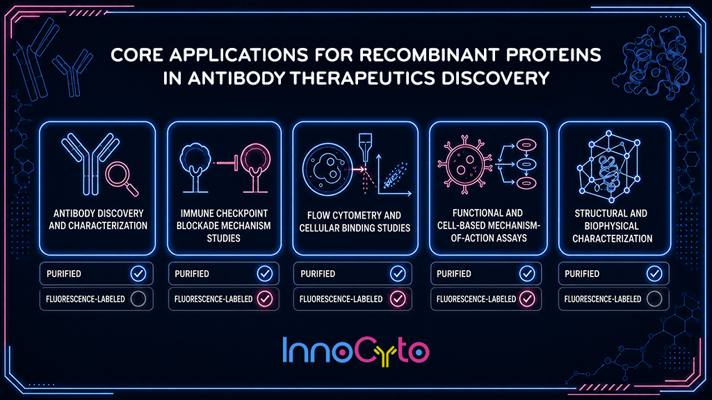
Recombinant proteins have become foundational reagents in the modern immuno-oncology laboratory. Where monoclonal antibodies serve as the therapeutic or detection endpoint, recombinant proteins, both purified and fluorescence-labeled, are increasingly the upstream tool that makes antibody discovery, functional characterization, and mechanistic research possible. InnoCyto's recombinant protein portfolio spans a broad set of cancer-relevant targets, including immune checkpoint receptors and ligands, cytokines, tumor-associated antigens, and Fc receptors. Because many of these proteins are themselves the direct targets of approved and investigational cancer immunotherapies, they occupy a unique dual role: as reagents for R&D and as molecular surrogates for the very biology that therapeutics are designed to modulate.
This technical note outlines the principal applications of InnoCyto’s recombinant proteins in cancer immunotherapy research, with attention to format-specific advantages (purified vs. fluorescence-labeled) and practical guidance for assay design.
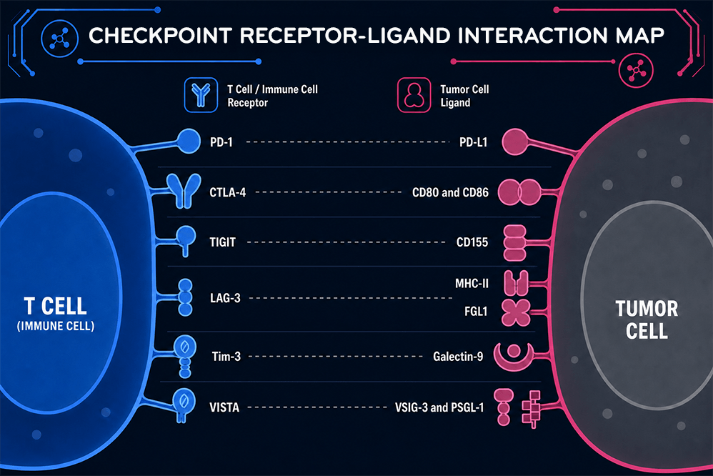
Cancer immunotherapy hinges on precisely modulating protein-protein interactions at the interface between tumor cells, antigen-presenting cells, and effector lymphocytes. Checkpoint receptors such as PD-1, CTLA-4, LAG-3, TIM-3, and TIGIT engage their cognate ligands (PD-L1, PD-L2, CD80/CD86, galectin-9, CD155) to deliver inhibitory signals that tumors exploit for immune evasion. Recombinant, correctly folded, and functionally validated versions of these receptor-ligand pairs allow researchers to reconstruct these interactions in a controlled, cell-free or cell-based context, critical for antibody screening, epitope mapping, and mechanism-of-action studies without the variability introduced by primary cells or transfected cell lines alone.
Purified recombinant antigens are the backbone of hybridoma screening, phage/yeast display panning, and hit validation. InnoCyto's checkpoint and tumor-antigen proteins (e.g., PD-L1, PD-1, CD47, HER2, EGFR, CD19) support:
Because checkpoint receptor-ligand pairs (PD-1/PD-L1, CTLA-4/CD80-CD86, TIGIT/CD155, LAG-3/FGL1) are central to current and emerging therapeutics, recombinant versions enable:
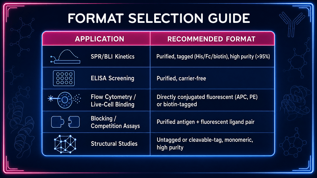
Fluorescence-labeled recombinant proteins (e.g., PD-L1-APC, PD-1-PE, CD155-FITC) allow direct visualization of receptor expression and ligand engagement on live cells without requiring a secondary detection antibody:
Recombinant cytokines, Fc receptors, and costimulatory ligands support functional immune assays beyond simple binding:
Purified proteins with high purity and defined oligomeric state support crystallography, cryo-EM, and computational modeling efforts used in structure-guided antibody and bispecific engineering, particularly relevant for novel checkpoint combinations where epitope geometry determines whether simultaneous dual binding is sterically feasible.
Recombinant proteins occupy a distinctive position in cancer immunotherapy research: they are simultaneously the molecular targets that define modern immuno-oncology and the enabling reagents that make studying those targets possible. From hybridoma screening and epitope mapping through kinetic characterization, flow cytometry-based functional assays, and structural biology, InnoCyto's purified and fluorescence-labeled recombinant proteins provide the consistency, purity, and format flexibility that discovery and validation workflows demand.
As the field moves toward increasingly sophisticated modalities, bispecific and trispecific antibodies, dual-checkpoint blockade, and engineered Fc-optimized therapeutics, the need for well-characterized, orthology-matched, and application-ready recombinant reagents will only grow. By offering both purified and fluorescently labeled formats across a broad panel of cancer- relevant targets, InnoCyto supports researchers at every stage of this pipeline, from first binding screen to final mechanistic confirmation, helping accelerate the path from target biology to clinical candidate.
Contact us for expert support.